What is Thermography?
Thermography or DITI (Digital Infrared Thermal Imaging) is an affordable and totally non-invasive, painless screening with no radiation and no contact with the body.
What is Thermography used for?
- To help in determining the cause of pain.
- To aid in the early detection of disease and pathology
- To evaluate sensory-nerve irritation or significant soft-tissue injury
- To define a previously diagnosed injury or condition
- To identify an abnormal area for further diagnostic testing
- To follow progress of healing and rehabilitation.
Breast Thermography
What is Breast Thermography?

Breast thermography is a 15 minute non invasive test of physiology. It is a valuable procedure for alerting your doctor to changes that can indicate early stage breast disease.
DITI’s role in breast health is to help in early detection and monitoring of abnormal physiology and the establishment of risk factors for the development or existence of pathology, whether benign or malignant. When used adjunctively with other procedures the best possible evaluation of breast health is made.
Thermography can detect the subtle physiologic changes that accompany breast pathology, whether it is cancer, fibrocystic disease, an infection or a vascular disease. Your doctor can then plan accordingly and lay out a careful program to further diagnose and /or MONITOR you during and after any treatment.
Angiogenisis

Angiogenesis is the physiological process through which new blood vessels form from pre-existing vessels. It is necessary to sustain the growth of a tumor. Breast thermography may be the first signal that such a possibility is developing.
Why is angiogenesis important in cancer? Angiogenesis plays a critical role in the growth of cancer because solid tumors need a blood supply if they are to grow beyond a few millimeters in size. Tumors can actually cause this blood supply to form by giving off chemical signals that stimulate angiogenesis.
Infrared is completely non-invasive, does not use radiation, does not compress the breast and is 100% safe.
Who should have this test?
Breast Screening Questions and Answers
Breast Screening Questions and Answers

All women can benefit from DITI breast screening, particularly younger women (30 - 50) with denser breast tissue.
Approximately 15% of all breast cancers occur in women under 45.
It takes years for a tumor to grow thus the earliest possible indication of abnormality is needed to allow for the earliest possible treatment and intervention. Thermography's role in monitoring breast health is to help in early detection and monitoring of abnormal physiology.
The faster a malignant tumor grows, the more Infrared radiation it generates. For younger women in particular, results from thermography screening can lead to earlier detection and, ultimately, longer life.
75% of women who get breast cancer have no family history of the disease.
Regardless of your family history, if a thermogram is abnormal you run a future risk of breast cancer that is 10 times higher than a first order family history of the disease. Thermography has the ability to provide women with a future risk assessment. If discovered, certain thermographic risk markers can warn a woman that she needs to work closely with her doctor with regular checkups to monitor her breast health.
Canadian researchers recently found that infrared imaging of breast cancers could detect minute temperature variations related to blood flow and demonstrate abnormal patterns associated with the progression of tumors. These images or thermograms of the breast were positive for 83% of breast cancers compared to 61% for clinical breast examination alone and 84% for mammography.
It's your body, it's your decision.
Breast Screening Questions and Answers
Breast Screening Questions and Answers
Breast Screening Questions and Answers

Why is thermal imaging useful for breast imaging?
Canadian researchers recently confirmed that infrared imaging of breast cancers could detect minute temperature variations related to blood flow and demonstrate abnormal patterns associated with the progression of tumors. These images, or thermograms of the breast, were positive for 83% of breast cancers compared to 61% for clinical breast examination alone and 84% for mammography. The 84% sensitivity rate of mammography alone was increased to 95% when infrared imaging was added.
Does it hurt to have a scan taken?
No. There is no contact with the body or painful breast compression.
If I have a suspicious mammogram or find a lump in a breast, should I have a thermogram?
Yes. The information provided by a thermography study can contribute useful additional information which ultimately helps your doctor with case management decisions. It is also important to establish a baseline for future comparison in order to monitor changes and the progress of any treatment.
Have clinical tests been done on thermal imaging?
Yes! Over 800 peer-reviewed studies on breast thermography exist in the index medicus literature. In this database, well over 300,000 women have been included as study participants. The numbers of participants in many studies are very large (10,000, 37,000, 60,000, 85,000, etc.) Some of these studies have followed patients for up to 12 years.
These clinical trials have demonstrated that breast thermography:
- Detects the first signs of a cancer up to 10 years before any other procedure can detect it.
- Significantly augments the long-term survival rates of its recipients by as much as 61%.
- When used as part of a multimodal approach (clinical examination + mammography + ultrasound + thermography), will detect 95% of early stage cancers.
Subscribe
Sign up to hear from us about specials, sales & deals.
Contact Us
Schedule your appointment today!
Hours
By appointment only.
Monday - Friday: 9am - 5pm
Sunday: Closed
Office in Coral Gables
Services
Full Body Thermography for Men & Women
30+ images of the whole body. Views included: head & neck, breasts, chest, abdomen and back, legs, arms, hands & feet
One Region of Interest for Men & Women
Choose from Head & Neck, Breast, Abdomen, Legs, Arms, Hands or Feet
Two Regions of Interest for Men & Women
Initial Breast Thermography
Your first breast screening
Three month follow-up Breast Thermography
Essential for establishing your thermal baseline prior to commencing annual screenings
Woman's Wellness Screening
Head & Neck, Breast & Abdomen
We accept cash, checks & most major credit cards.


More info
Who takes the images?
Overview of Thermography
Overview of Thermography

Hi! My name is Jennifer Kaufman and I am a graduate of NYU, a ACCT certified clinical thermographer and Dr. Sears certified Health Coach. In 2014 I came across an article titled Thermography vs Mammography. At the time I did not know it would change my life. I did a full body thermography screening and discovered I had a lot of inflammation. Fortunately for me I had no serious issues but clearly thermography was telling me to change my lifestyle before I developed symptoms. So I did! I changed my eating habits, I changed my exercise habits and I completely changed my understanding of health. Within a month of my initial screening I decided to become a thermographer. It is a challenge to be healthy in today's world. I hope I have the opportunity to share this amazing FDA approved medical screening along with my experience and knowledge with you.
Overview of Thermography
Overview of Thermography
Overview of Thermography
Click on Image for more info.
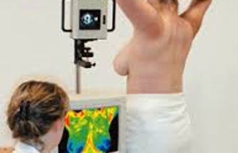
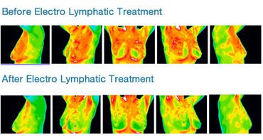
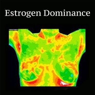
Medical DITI is a noninvasive diagnostic technique that allows the examiner to visualise and quantify changes in skin surface temperature. An infrared scanning device is used to convert infrared radiation emitted from the skin surface into electrical impulses that are visualised in colour on a monitor. This visual image graphically maps the body temperature and is referred to as a thermogram. The spectrum of colours indicate an increase or decrease in the amount of infrared radiation being emitted from the body surface. Since there is a high degree of thermal symmetry in the normal body, subtle abnormal temperature asymetry's can be easily identified.
Most patients use thermography for breast imaging but it can also be used to help in determining the cause of pain. It can be used in the early detection of temperature change, to evaluate sensory-nerve irritation or significant soft-tissue injury, to help define a previously diagnosed injury or condition, or to identify an abnormal area for further diagnostic testing. Thermography is an adjunctive tool and provides a different set of results to other tests, allowing a healthcare provider to have a wider set of information to aid diagnosis and treatment. There are conditions or diseases for which a different test would be more appropriate, but your nearest thermography clinic should be able to advise accordingly.
What to expect.
Overview of Thermography
Preparation for Thermography
A thermal scan takes approximately 10 to 45 minutes depending on which part of the body is being scanned. You will remove all jewelry and clothes from the part of the body being scanned (for full body scans you leave underpants on). For a breast scan, you will be ask to disrobe from the waist up.
You will need to sit for a few minutes while your skin is equalizing with the room temperature. At that time we will go over your paper work.
Preparation for Thermography
Who interprets my images and writes the report?
Preparation for Thermography
There are a few guidelines for preparing for a thermal scan:
- Do not have physical therapy, massage, or electromyography on the same day thermography is performed.
- Do not participate in vigorous exercise 2 hours prior to the test.
- Do not smoke for 2 hours before the test.
- Do not use lotions, deodorants, powder or antiperspirant or shave on day of test.
- Stay out of strong sunlight on the day of test.
- Do not have body work, lymphatic drainage, or acupuncture 3 days prior.
- Wait 3 months post surgery, radiation therapy, and chemotherapy.
- Wait 3 months post lactation.
- There are no dietary or medication restrictions on the day of your scan but no excessive hot or cold drinks prior to the test.
Who interprets my images and writes the report?
Who interprets my images and writes the report?
Who interprets my images and writes the report?
Medical doctors or themologists who represent a variety of specialties such as Emergency Medicine, Functional Medicine, OB/GYN, General Practice, and Radiology interpret the thermal images for abnormal heat patterns and asymmetries. Board certification and training is provided by the American College of Clinical Thermology (ACCT). The report is usually ready within 3-5 business days.
Download
Please download the applicable forms below prior to your appointment.
- Patient Preparation Sheet
- Patient Intake Form
- Review of Systems
- Breast Questionnaire (for women only)
- EMI Authorization
- HIPAA Form
- Upper Body Questionnaire (for half body screening only)
- Full Body Questionnaire (for full body screening only)
Patient Preparation Sheet (pdf)
DownloadPatient Intake Form (pdf)
DownloadReview of Systems (pdf)
DownloadBreast-Questionnaire (pdf)
DownloadEMI Authorization (pdf)
DownloadHIPAA Form (pdf)
DownloadFull Body Questionnaire (pdf)
DownloadPatient Preparation sheet general (pdf)
DownloadUpper Body Questionnaire (pdf)
Download
